





Diagnostics & Academic Works
Clinical Portfolio
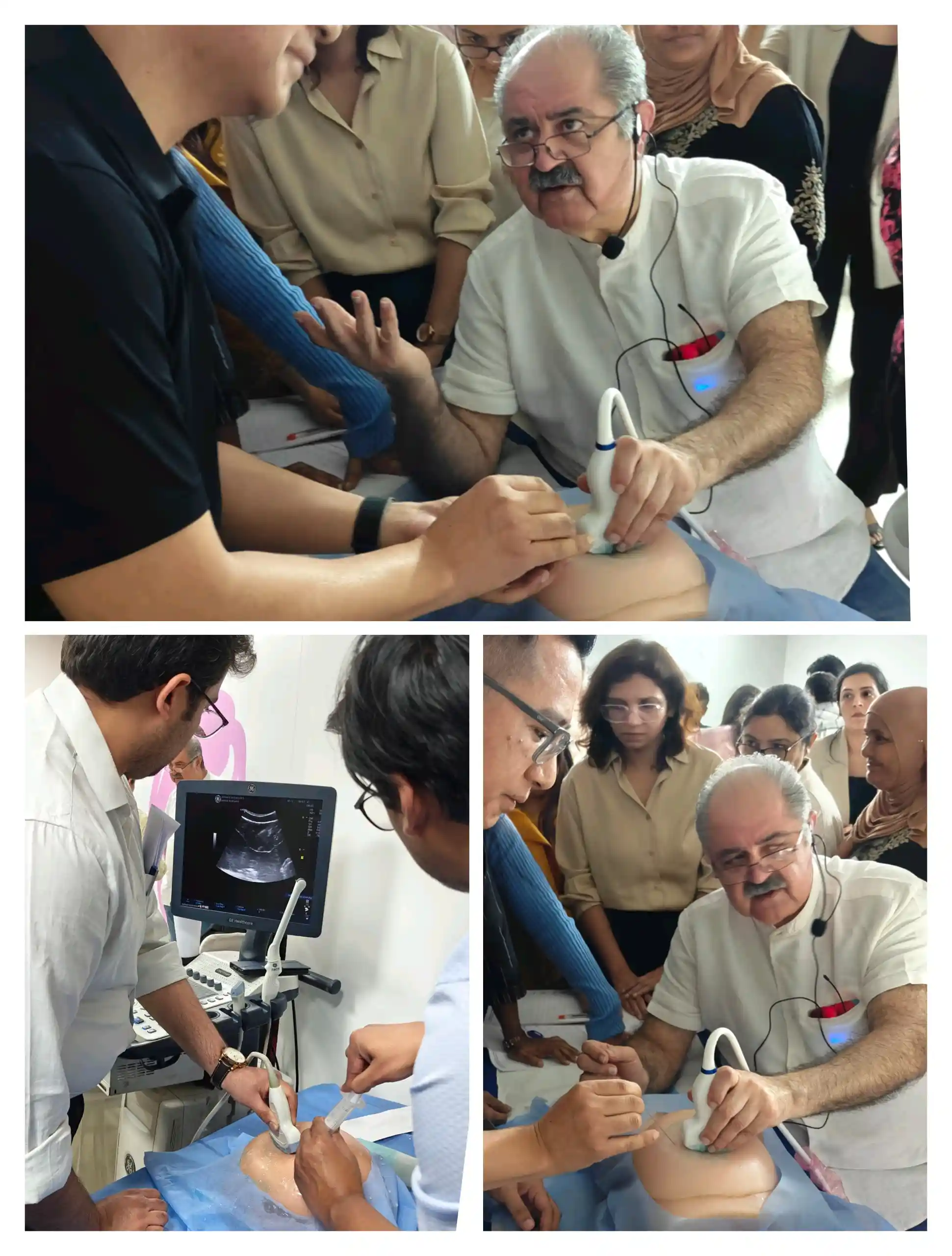
A comprehensive reference library of clinical cases, ultrasound-guided procedures, and professional academic lecture credentials.
.webp)
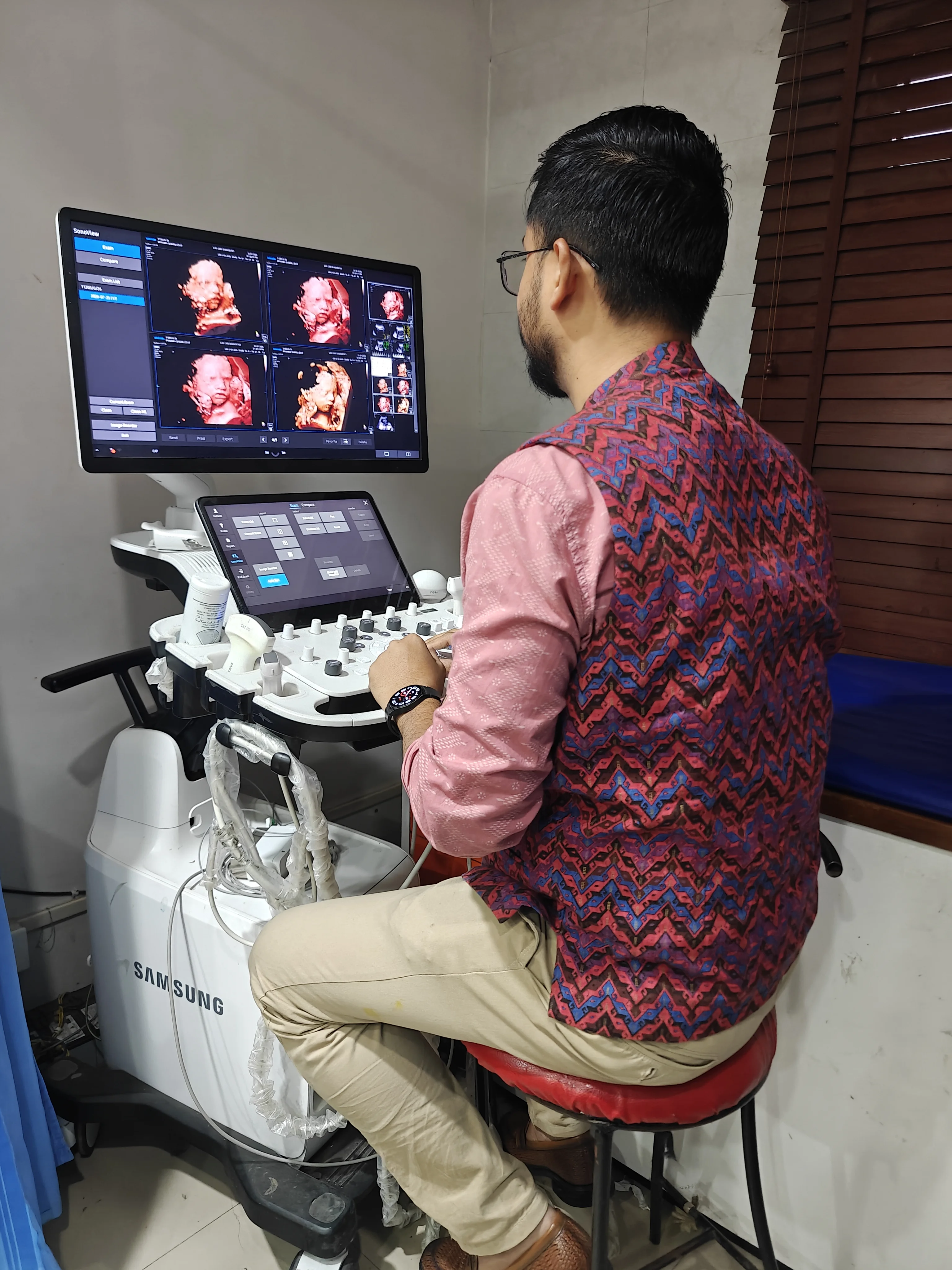














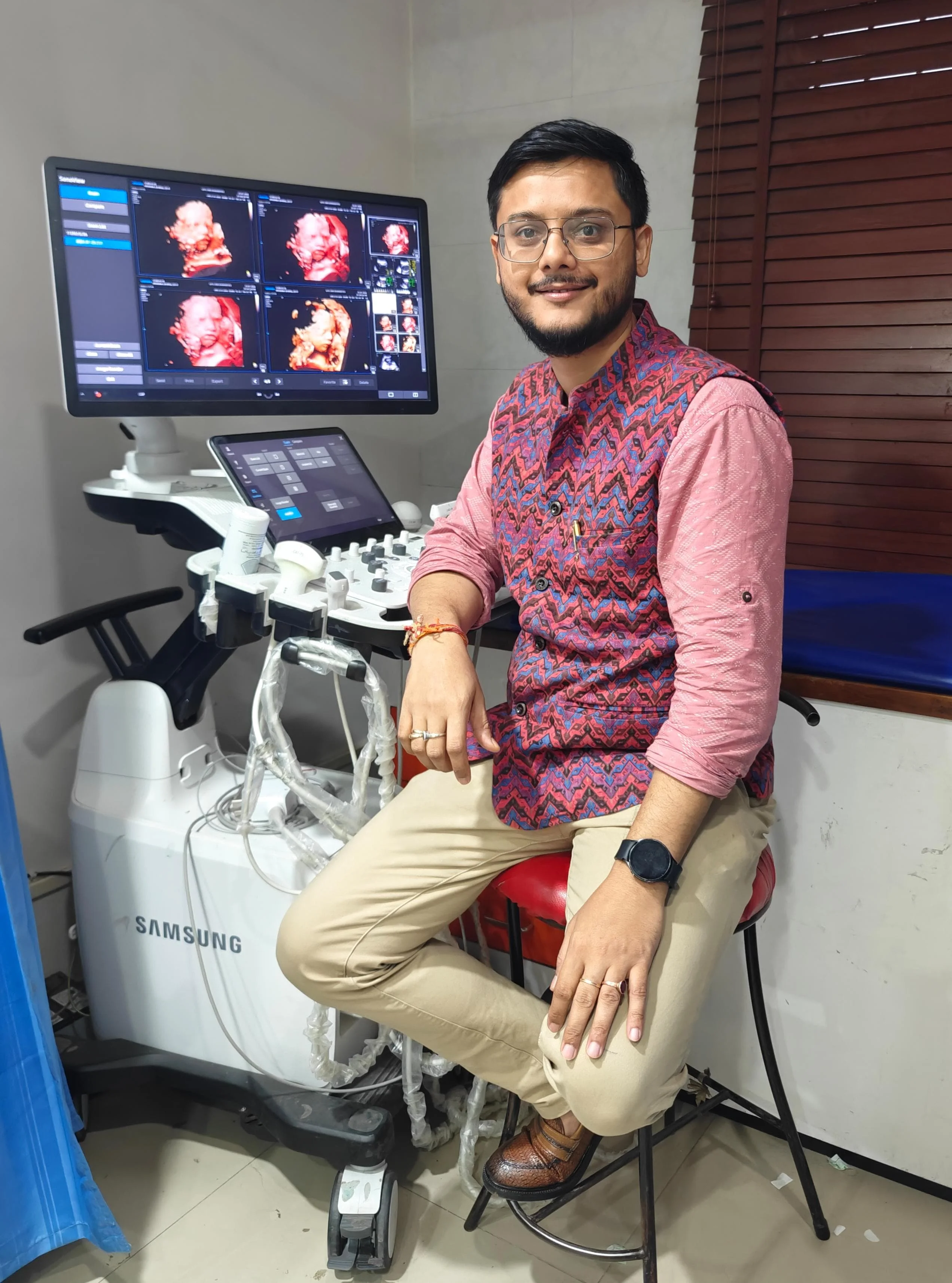
Consultant Radiologist & Fetal Medicine Specialist
Precision Fetal Medicine Scans & Expert Radiology Reports
"We don't rush examinations. We take the time needed to deliver accurate, complete, and clinically meaningful imaging."
PATIENT CARE MANDATE"Excellence in fetal imaging begins with patience, precision, and attention to every detail."
FETAL MEDICINE PHILOSOPHY
"A radiology report should be more than a description of findings—it should answer the clinician's question, guide patient management, and add genuine value to clinical care."
I completed my MBBS from Assam Medical College & Hospital (AMCH), Dibrugarh, followed by my MD in Radiodiagnosis from Gauhati Medical College & Hospital (GMCH), Guwahati. Driven by the pursuit of medical excellence, I further earned the title of Diplomate of National Board (DNB) in Radiodiagnosis, validating my expertise across all major modalities.
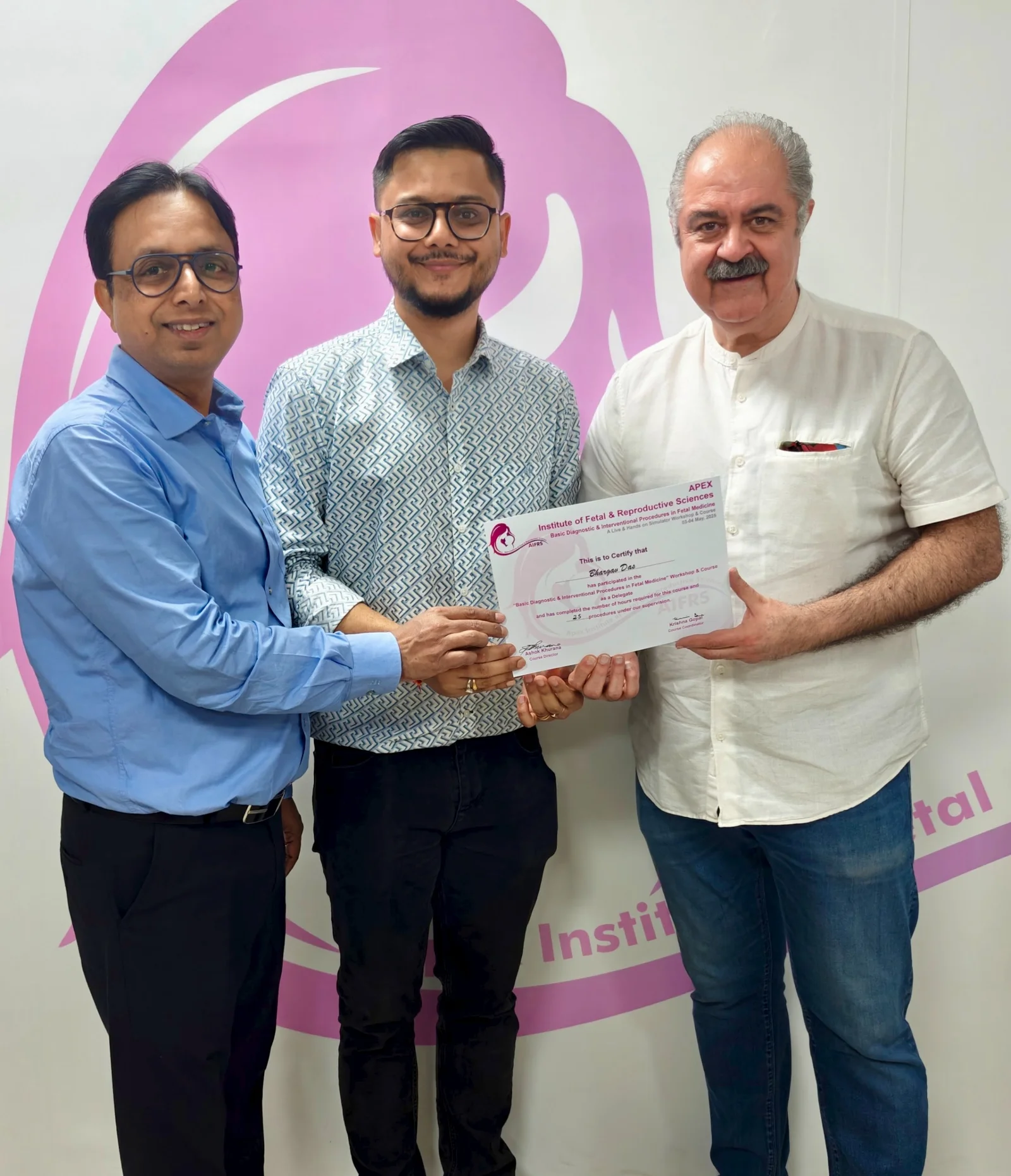
Recognizing the critical importance of early prenatal diagnostics, I pursued advanced training in Fetal Medicine through Scholar MD. To further refine my subspecialist skills, I completed a dedicated observership under the legendary fetal medicine pioneer Dr. Bimal Sahani and received specialized hands-on training in advanced fetal interventions from Dr. Ashok Khurana.
Today, I specialize in Fetal Medicine, Obstetric & Gynecological Imaging, Doppler Studies, Musculoskeletal (MSK) Ultrasound, and Cross-sectional Imaging (CT). I remain committed to continuous learning and updating my practices to international standards.
Assam Medical College & Hospital (AMCH), Dibrugarh
Gauhati Medical College & Hospital (GMCH), Guwahati
National Board of Examinations
Scholar MD, Training under Dr. Bimal Sahani & Dr. Ashok Khurana
Guiding principles behind my diagnostic radiology and fetal medicine practice
I believe that radiology is far more than interpreting images—it is an integral part of patient care. Every radiology report should answer the clinician's question, guide patient management, and provide meaningful clinical insight rather than simply describe imaging findings.
My approach combines precision imaging, evidence-based reporting, and clear communication to support informed clinical decision-making and improve patient outcomes. Every examination is performed with meticulous attention to detail, compassion, and an unwavering commitment to diagnostic excellence.
Every patient who undergoes an imaging examination is seeking answers, reassurance, and hope. Every referring clinician depends on imaging to make timely and accurate decisions. This responsibility inspires me to pursue the highest standards of diagnostic accuracy while continually refining my knowledge and skills.
My passion lies in Fetal Medicine, Musculoskeletal Ultrasound, and Ultrasound-Guided Interventional Procedures, where imaging has a direct impact on patient care and clinical outcomes. Beyond clinical practice, I am deeply committed to lifelong learning, academic excellence, and sharing knowledge with fellow healthcare professionals and future radiologists.
I am committed to delivering high-quality, evidence-based diagnostic imaging with integrity, precision, and professionalism. Every patient deserves careful attention, and every referring clinician deserves a report that is clear, clinically relevant, and actionable.
I strive to maintain international standards in radiology through continuous education, adoption of evolving imaging techniques, and close collaboration with clinicians. My goal is to build lasting trust by providing reliable diagnostic expertise, personalized patient care, and meaningful contributions to medical education and research.
Offering advanced diagnostic insight across multiple imaging disciplines
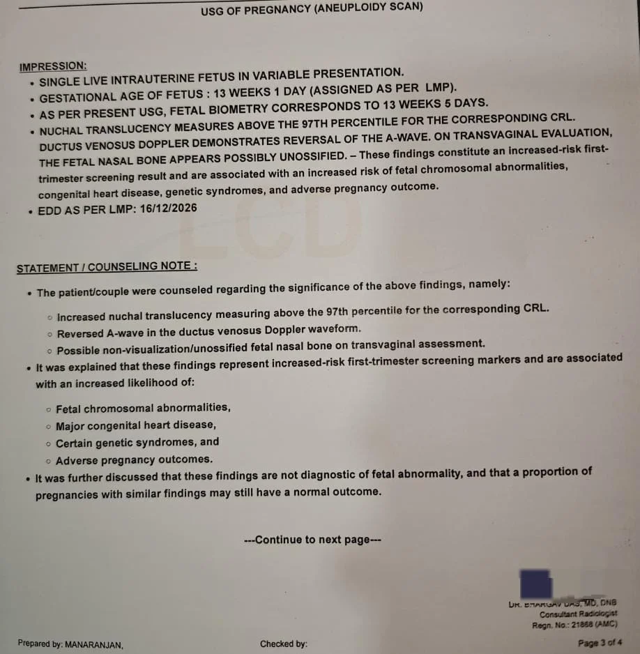
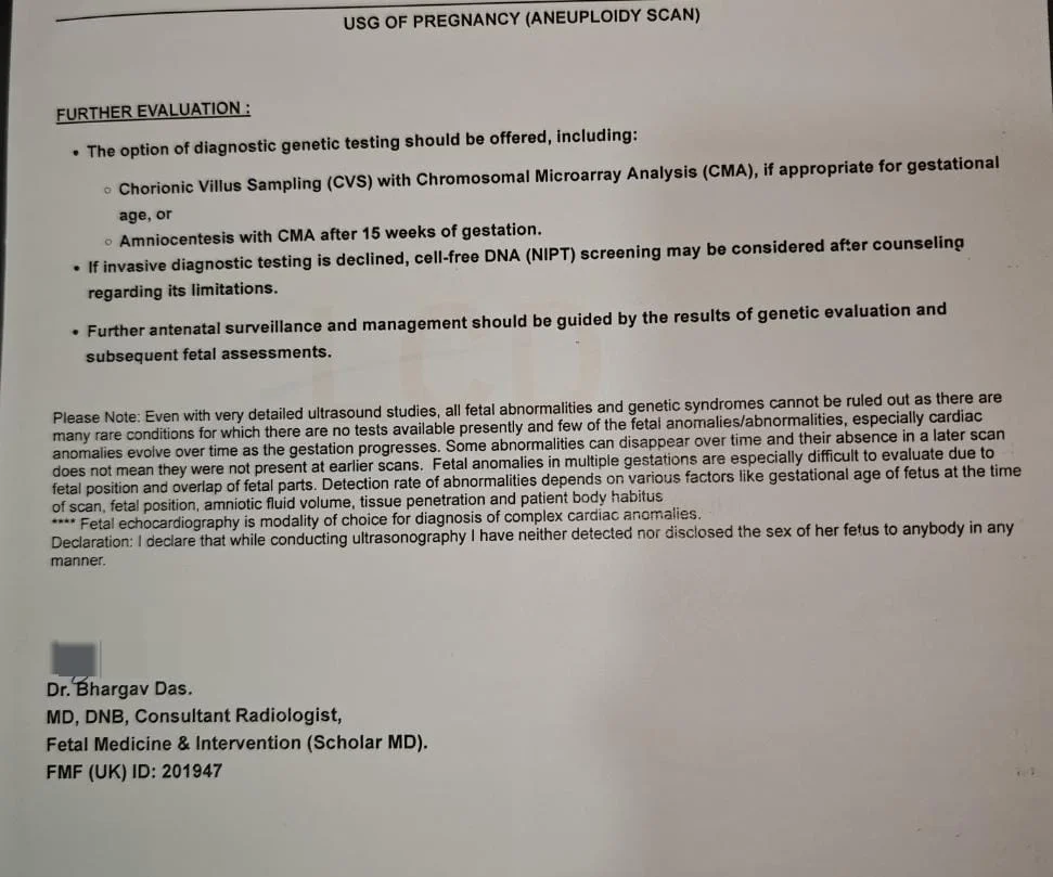
Specialized early pregnancy assessments, NT scans, Level I & Level II detailed anomaly scans to monitor fetal development and detect abnormalities early.
High-resolution pelvic and transvaginal ultrasound scans to diagnose structural issues, monitor pregnancy progress, and investigate gynecological health problems.
Advanced vascular flow assessments including fetal Doppler, renal Doppler, and peripheral vascular studies to evaluate circulations and detect vascular compromises.
Dynamic and static high-frequency imaging of soft tissues, muscles, ligaments, nerves, and joints to check for tears, sprains, and inflammatory conditions.
Expert diagnostic interpretation of CT scans for brain, chest, abdomen, pelvis, and musculoskeletal systems, delivering detailed anatomical breakdowns.
Safe, minimally invasive ultrasound-guided procedures including FNAC, core needle biopsies, joint injections, cyst aspirations, and fluid drainage.
Genuine feedback from patients receiving specialized fetal medicine scans, diagnostic imaging, and ultrasound-guided procedures.
"Dr. Bhargav Das is extremely patient and meticulous. During our Level II anomaly scan, he spent adequate time explaining every detail of our baby’s heart and spine anatomy. He reassured us completely and never rushed the examination."
"We were advised a follow-up scan for a soft marker detected elsewhere. Dr. Bhargav conducted a thorough 3D fetal evaluation and explained clearly why it was a normal variation. His clinical depth and gentle bedside manner put our minds at ease immediately."
"I had persistent wrist pain that wasn't resolving. Dr. Bhargav performed a real-time MSK ultrasound and identified tenosynovitis with high accuracy. His detailed reporting helped my treating doctor prescribe the exact targeted care."
"During the early weeks of my pregnancy, I was filled with anxiety. Dr. Bhargav handled my viability scan with so much empathy and care. Hearing the heartbeat for the first time with his reassuring explanation was an unforgettable experience."
"Underwent an ultrasound-guided aspiration procedure with Dr. Bhargav. He was remarkably skilled and gentle—the procedure was completely swift and painless. High-quality diagnostic expertise!"
"A detailed 3D pelvic ultrasound examination for fertility evaluation. Dr. Bhargav's comprehensive reporting provided my gynecologist with exact clinical clarity for my treatment plan. Highly recommended radiologist!"
"Finding a radiologist who takes time to answer every question and explain diagnostic findings in simple words is rare. Dr. Bhargav is truly dedicated to patient care and accurate clinical reporting."
Comprehensive diagnostic guidance for patients, referring obstetricians, infertility specialists, and orthopaedic surgeons.
A viability scan is an early pregnancy ultrasound usually performed between 6 and 9 weeks of pregnancy. It confirms that the pregnancy is located within the uterus, determines the gestational age, detects fetal cardiac activity, and helps identify conditions such as early pregnancy failure or ectopic pregnancy.
Most women benefit from an early ultrasound between 6 and 9 weeks of pregnancy. If you experience pain, bleeding, have had previous miscarriages, fertility treatment, or uncertain pregnancy dates, your doctor may recommend an earlier scan.
The NT scan is not compulsory, but it is strongly recommended. Performed between 11 weeks and 13 weeks + 6 days, it provides valuable information about your baby's early development and helps assess the risk of certain chromosomal abnormalities when combined with appropriate screening tests.
NT Scan: Performed in the first trimester (11–13+6 weeks), focusing on early fetal anatomy and chromosomal risk assessment.
Anomaly (Level II) Scan: Performed between 18 and 24 weeks (ideally around 21 weeks), providing a comprehensive evaluation of your baby's organs and overall development.
The anomaly scan thoroughly examines your baby's vital structures:
It also evaluates overall fetal growth and anatomical well-being.
Not at all. In most cases, this is completely normal and does not mean there is a problem with your baby.
An anomaly scan is one of the most detailed ultrasound examinations performed during pregnancy. To evaluate every organ accurately, we need clear views of the baby's heart, brain, face, spine, kidneys, limbs, and other structures. The quality of the examination depends greatly on your baby's position. Babies often curl up, keep their hands in front of the face, turn away, or remain in positions that temporarily prevent us from obtaining required images.
During the scan, you may be asked to walk for a few minutes, change your position, or wait for some time before continuing. If the position remains unfavourable, returning on another day ensures every structure is evaluated properly without rushing.
“At our centre, we never rush an anomaly scan. If your baby's position does not allow complete evaluation, we prefer to repeat the examination rather than provide an incomplete assessment. Our priority is accuracy—not speed.”
No. Most isolated soft markers are found in completely healthy babies. Their significance depends on maternal age, previous screening results, and whether other ultrasound findings are present.
Yes. Diagnostic ultrasound has been used safely during pregnancy for several decades when performed appropriately by trained medical professionals for clinical indications.
A repeat scan may be recommended because:
Repeat imaging significantly enhances diagnostic accuracy.
MSK ultrasound evaluates soft tissue structures including muscles, tendons, ligaments, joints, nerves, and soft tissue swellings. It is commonly used for sports injuries, tendon disorders, ganglion cysts, arthritis, and nerve entrapments.
These procedures are generally well tolerated and performed under local anaesthesia. Real-time ultrasound guidance maximizes precision, minimizes discomfort, and ensures safety.
Patients presenting with vaginal bleeding, pelvic pain, previous miscarriages, IVF pregnancies, uncertain dates, suspected ectopic pregnancy, or pregnancy of unknown location benefit from early ultrasound assessment.
Diagnosis strictly follows internationally accepted ultrasound criteria to avoid false-positive conclusions. When findings are equivocal, repeat ultrasound after an appropriate interval is recommended before confirming failure.
All pregnant women should be offered an NT scan between 11 and 13+6 weeks. It is particularly valuable in cases of advanced maternal age, positive screening tests, previous aneuploidy, IVF pregnancies, and high-risk pregnancies.
Early anomaly assessment is valuable in increased NT, high-risk pregnancies, maternal diabetes, IVF conception, previous fetal anomalies, monochorionic twins, positive serum screening, and suspected structural malformations.
Soft markers are evaluated alongside maternal age, biochemical screening, presence of additional anomalies, and established international guidelines. Isolated markers carry significantly different risk profiles compared to multiple associated markers.
Referral is indicated for increased NT, abnormal 4-chamber or outflow tract views, maternal diabetes, IVF pregnancies, family history of congenital heart disease, monochorionic twins, or abnormal screening results.
Doppler studies are recommended in fetal growth restriction (FGR), hypertensive disorders, suspected fetal anaemia, high-risk pregnancies, twin gestations, and maternal vascular disorders.
Yes. Reports follow a systematic organ-based approach with clinically relevant impressions, differential diagnoses where appropriate, and clear management recommendations.
Answering clinicians directly is our priority. Referring clinicians are welcome to contact Dr. Bhargav to discuss complex cases, clarify imaging nuances, or coordinate optimal patient management.
Yes. Targeted neurosonography, serial follow-up examinations, growth surveillance, and Doppler assessment are performed whenever clinically indicated.
A comprehensive infertility scan evaluates uterine architecture, endometrial thickness and pattern, ovarian morphology, Antral Follicle Count (AFC), pelvic organ mobility, congenital uterine anomalies, and endometrial pathology.
3D TVS is essential for evaluating Müllerian anomalies (septate vs. bicornuate uterus), endometrial polyps, submucosal fibroids, intrauterine synechiae, infertility work-ups, and recurrent pregnancy loss.
Yes. High-resolution TVS accurately identifies features of adenomyosis and endometriomas, and can detect deep infiltrating pelvic endometriosis when performed systematically.
Yes. Antral Follicle Count (AFC) measured via transvaginal ultrasound is a key biomarker of ovarian reserve and is routinely evaluated during infertility protocols.
MSK ultrasound is often preferred as a first-line modality for superficial tendon disorders, ligament tears, ganglion cysts, bursitis, dynamic tendon subluxation, nerve entrapments, and real-time image-guided interventions.
Dynamic ultrasound enables real-time examination during joint movement, allowing immediate evaluation of tendon snapping, subluxation, muscular hernias, and joint impingement that static MRI may miss.
Yes. High-frequency probes accurately characterize partial vs. complete tendon tears, tendinosis, calcific tendinopathy, and tenosynovitis.
Yes. High-resolution ultrasound reliably distinguishes fluid-filled cystic lesions from solid vascularized masses and maps their relation to adjacent joint capsules and nerve branches.
Aspiration is indicated for symptomatic cysts causing pain, nerve compression, or functional impairment following clinical evaluation.
Yes. High-frequency MSK ultrasound measures cross-sectional nerve swelling in median (carpal tunnel), ulnar (cubital tunnel), radial, tibial, and peroneal nerve entrapments.
Yes. Power Doppler USG detects early subclinical synovitis, tenosynovitis, joint effusions, and bone erosions before radiographic changes appear.
Yes. Real-time guidance ensures target accuracy for joint aspirations, bursa drainage, targeted therapeutic injections, and soft tissue biopsies.
An NT scan is strictly performed between 11 weeks and 13 weeks + 6 days (fetal CRL between 45mm and 84mm).
An anomaly scan evaluates dozens of microscopic fetal structures. If your baby is curled up or facing backwards, extra time or maternal walking is needed to ensure every organ is thoroughly examined.
Returning for a repeat scan is done to assess structures temporarily hidden by fetal positioning. It reflects our commitment to thoroughness, not a sign of abnormality.
Soft markers are non-specific anatomical variations that are frequently seen in completely normal babies. Isolated markers rarely indicate underlying chromosomal problems.
Fetal echocardiography is ideally performed between 18 and 24 weeks when cardiac structures or screening tests warrant detailed evaluation.
While high-resolution ultrasound detects a vast majority of major structural anomalies, certain functional or micro-structural conditions may not manifest visually on ultrasound.
Yes, 3D coronal volume reconstruction provides gold-standard diagnostic precision for uterine anomalies like septate or bicornuate uterus.
High-resolution TVS accurately diagnoses ovarian endometriomas and deep infiltrating pelvic endometriosis.
MSK ultrasound offers superior spatial resolution for superficial tendons and allows dynamic movement evaluation that MRI cannot replicate.
Yes. Ultrasound confirms cystic nature, internal acoustic enhancement, and joint neck origin with high diagnostic certainty.
Ultrasound-guided FNAC is indicated for palpable or incidental soft tissue, thyroid, or lymph node lesions requiring definitive cytological diagnosis.
Umbilical and middle cerebral artery (MCA) Doppler guides timing of delivery by monitoring placental resistance and fetal blood flow redistribution.
Precision diagnostic scans and ultrasound-guided procedures of international standards
Committed to accurate diagnosis, advanced imaging, and personalized patient care using state-of-the-art ultrasound and CT technology.
Get in touch directly for appointments, reports lookup, or professional queries
+91 9957510876
Message or call directly for appointments and imaging queries
Email for official, academic, or professional collaboration
Follow Dr. Bhargav Das across official platforms & Google Business Profile
Available for clinical diagnostic scans at the following centers. Call the clinic desk directly to schedule your scan.
Maligaon Branch
Baihata Branch
A comprehensive reference library of clinical cases, ultrasound-guided procedures, and professional academic lecture credentials.
Evidence-based medical articles, fetal diagnostics insights, and clinical ultrasound guidance by Dr. Bhargav Das
The first few weeks of pregnancy are often filled with excitement and anxiety. A viability scan, usually performed between 6 and 9 weeks of pregnancy, is one of the most important ultrasound examinations during this period...
The first few weeks of pregnancy are often filled with excitement and anxiety. A viability scan, usually performed between 6 and 9 weeks of pregnancy, is one of the most important ultrasound examinations during this period.
The scan confirms whether the pregnancy is developing normally by identifying the gestational sac, yolk sac, embryo, and fetal heartbeat. It also helps determine the exact gestational age and excludes conditions such as ectopic pregnancy or early pregnancy failure.
Early pregnancy failure occurs when the pregnancy does not progress as expected. However, it is important to remember that a single early ultrasound may not always provide a definitive answer. In many cases, a follow-up scan after 7–14 days is necessary before making a diagnosis.
Accurate diagnosis requires adherence to internationally accepted ultrasound criteria to avoid misdiagnosis of a potentially viable pregnancy.
Never rush to conclusions after an early scan. Proper timing and expert ultrasound assessment are essential for safe and accurate diagnosis.
The Nuchal Translucency (NT) Scan is performed between 11 weeks and 13 weeks + 6 days of pregnancy. It is one of the most valuable screening tests in early pregnancy...
The Nuchal Translucency (NT) Scan is performed between 11 weeks and 13 weeks + 6 days of pregnancy. It is one of the most valuable screening tests in early pregnancy.
The scan measures the fluid-filled space behind the baby's neck and evaluates several important anatomical markers. Combined with maternal age and blood tests, it helps estimate the risk of chromosomal conditions such as Down syndrome.
Beyond screening for chromosomal abnormalities, the NT scan also assesses early fetal anatomy, confirms fetal viability, determines gestational age, and identifies multiple pregnancies.
An increased NT measurement does not necessarily mean that the baby has a genetic disorder, but it may indicate the need for further evaluation and counselling.
The NT scan is much more than measuring a single number—it provides an early and comprehensive assessment of your baby's development.
The Anomaly Scan, also known as the Level II Scan, is one of the most important ultrasound examinations during pregnancy, ideally performed around 20–22 weeks for comprehensive evaluation...
The Anomaly Scan, also known as the Level II Scan, is one of the most important ultrasound examinations during pregnancy. It can be performed between 18 and 24 weeks of gestation, although the ideal time is around 20–22 weeks, with approximately 21 weeks often providing the best opportunity for a comprehensive evaluation.
This detailed examination carefully assesses your baby's major organ systems, including the brain, face, spine, heart, lungs, diaphragm, abdomen, stomach, kidneys, urinary bladder, limbs, hands, feet, placenta, umbilical cord, and amniotic fluid. The scan also evaluates fetal growth and overall well-being.
One important aspect that many parents are unaware of is that the quality of the examination depends significantly on the baby's position. Babies frequently change position, and certain structures may not be clearly visible during a single examination. To obtain the best possible images, you may be asked to walk around, change your position, or wait for some time before the scan is continued. In some cases, a repeat visit on another day may be necessary to complete the examination. This is a normal part of high-quality fetal imaging and should not be interpreted as a cause for concern.
The primary aim of the anomaly scan is to identify structural abnormalities, assess normal fetal development, and detect conditions that may require further evaluation, counselling, specialised pregnancy care, or treatment after birth. Fortunately, the majority of anomaly scans are reassuring and demonstrate normal fetal development.
The anomaly scan is much more than a routine ultrasound. Performed at the right stage of pregnancy and with careful attention to fetal position, it provides one of the most comprehensive assessments of your baby's growth, anatomy, and well-being. Sometimes, patience and repeat imaging are essential to ensure that every important structure is evaluated thoroughly.
A soft marker is a subtle ultrasound finding that, by itself, is usually not a birth defect. These markers may be seen in completely healthy babies but can sometimes be associated with increased risk...
A soft marker is a subtle ultrasound finding that, by itself, is usually not a birth defect. These markers may be seen in completely healthy babies but can sometimes be associated with an increased risk of certain chromosomal conditions.
Examples include:
The significance of a soft marker depends on whether it is isolated or associated with other ultrasound findings, maternal age, and previous screening results.
In many pregnancies, isolated soft markers require no treatment and often resolve naturally before birth.
Finding a soft marker does not mean there is a problem with your baby. Each finding should be interpreted in the context of the entire pregnancy by an experienced fetal medicine specialist.
Müllerian anomalies are congenital abnormalities of the uterus that occur during fetal development. Many women remain unaware of these conditions until experiencing infertility or pregnancy loss...
Müllerian anomalies are congenital abnormalities of the uterus that occur during fetal development. Many women remain unaware of these conditions until they experience infertility, recurrent miscarriages, or pregnancy complications.
Common uterine anomalies include a septate uterus, bicornuate uterus, unicornuate uterus, and uterus didelphys. Accurate diagnosis is important because some anomalies, particularly a septate uterus, can be successfully treated to improve reproductive outcomes.
Ultrasound, especially 3D transvaginal ultrasound, has become the preferred first-line imaging modality for evaluating uterine anatomy. It provides excellent visualization of both the uterine cavity and external contour, helping differentiate between various Müllerian anomalies.
Early and accurate diagnosis of uterine anomalies allows appropriate counselling and management, improving the chances of a successful pregnancy.
Ultrasound plays a central role in the evaluation of infertility. A comprehensive fertility scan assesses the uterus, endometrium, ovaries, ovarian reserve, and pelvic anatomy...
Ultrasound plays a central role in the evaluation of infertility. A comprehensive fertility scan assesses the uterus, endometrium, ovaries, ovarian reserve, and pelvic anatomy while identifying conditions that may affect conception.
3D Transvaginal Ultrasound provides additional information by accurately evaluating the uterine cavity, detecting congenital uterine anomalies, submucosal fibroids, and endometrial polyps that may interfere with implantation.
Depending on the clinical situation, ultrasound can also monitor follicular development, ovulation, and endometrial receptivity during fertility treatment.
A detailed infertility ultrasound, particularly with 3D imaging when indicated, provides valuable information that helps guide fertility treatment and improve reproductive outcomes.
An ectopic pregnancy occurs when a fertilized egg implants outside the uterine cavity, most commonly within the fallopian tube. It is a potentially life-threatening condition if not diagnosed early...
An ectopic pregnancy occurs when a fertilized egg implants outside the uterine cavity, most commonly within the fallopian tube. It is a potentially life-threatening condition if not diagnosed early.
Women may present with delayed periods, abdominal pain, vaginal bleeding, or shoulder pain, although some have minimal symptoms.
Transvaginal ultrasound combined with serum beta-hCG plays a crucial role in early diagnosis. The scan looks for an intrauterine pregnancy, adnexal masses, and free fluid within the pelvis.
Early diagnosis allows timely medical or surgical treatment while reducing complications and preserving future fertility whenever possible.
Any woman with a positive pregnancy test and pain or bleeding should undergo prompt ultrasound evaluation to exclude an ectopic pregnancy.
Ovarian cysts are common and are often discovered during routine ultrasound examinations. Most are functional cysts related to the normal menstrual cycle and resolve without treatment...
Ovarian cysts are common and are often discovered during routine ultrasound examinations. Most are functional cysts related to the normal menstrual cycle and resolve without treatment.
Ultrasound helps determine the size, appearance, and internal characteristics of a cyst, allowing differentiation between simple cysts, haemorrhagic cysts, endometriomas, dermoid cysts, and cystic ovarian tumours.
Most simple ovarian cysts require only observation, while complex cysts or persistent lesions may need further evaluation or surgical consultation.
Most ovarian cysts are benign. A detailed ultrasound examination helps determine whether observation or further treatment is required.
Polycystic Ovary Syndrome (PCOS) is one of the most common hormonal disorders affecting women of reproductive age. Symptoms may include irregular periods, acne, and difficulty conceiving...
Polycystic Ovary Syndrome (PCOS) is one of the most common hormonal disorders affecting women of reproductive age. Symptoms may include irregular periods, acne, excessive hair growth, difficulty conceiving, and weight gain.
Ultrasound may demonstrate enlarged ovaries containing multiple small follicles arranged around the periphery. However, PCOS cannot be diagnosed by ultrasound alone. Diagnosis is based on clinical findings, hormonal evaluation, and ultrasound features using accepted diagnostic criteria.
Lifestyle modification, weight management, and appropriate medical treatment can significantly improve symptoms and fertility outcomes.
The presence of polycystic ovaries on ultrasound does not automatically mean a woman has PCOS. Clinical assessment remains equally important.
Endometrial polyps are localized overgrowths of the lining of the uterus. They commonly present with abnormal uterine bleeding, intermenstrual spotting, or infertility...
Endometrial polyps are localized overgrowths of the lining of the uterus. They commonly present with abnormal uterine bleeding, intermenstrual spotting, or infertility, although many women have no symptoms.
Transvaginal ultrasound is often the first investigation and may identify a focal lesion within the endometrial cavity. Colour Doppler may demonstrate a characteristic single feeding vessel, increasing diagnostic confidence. In selected cases, saline infusion sonography or hysteroscopy may be recommended for further evaluation.
Removal of symptomatic polyps may improve bleeding symptoms and enhance fertility in appropriately selected patients.
Endometrial polyps are usually benign and readily diagnosed with ultrasound. Early identification helps guide appropriate treatment, particularly in women with infertility or abnormal uterine bleeding.
A ganglion cyst is a common, non-cancerous fluid-filled swelling that usually develops around the wrist, hand, ankle, or foot. Musculoskeletal Ultrasound (MSK USG) is an excellent imaging tool...
A ganglion cyst is a common, non-cancerous fluid-filled swelling that usually develops around the wrist, hand, ankle, or foot.
Musculoskeletal Ultrasound (MSK USG) is an excellent imaging tool for evaluating ganglion cysts. It accurately identifies the cyst, determines its size, confirms its fluid nature, and demonstrates its relationship with nearby tendons, joints, and nerves.
Ultrasound also helps differentiate ganglion cysts from solid soft tissue masses that may require different treatment.
In selected patients, ultrasound can guide needle aspiration safely and accurately.
MSK ultrasound provides a quick, painless, radiation-free, and highly accurate assessment of ganglion cysts while also assisting in image-guided treatment when appropriate.
Tenosynovitis refers to inflammation of the tendon sheath and is a common cause of pain, swelling, and restricted movement around joints. MSK ultrasound allows dynamic real-time evaluation...
Tenosynovitis refers to inflammation of the tendon sheath and is a common cause of pain, swelling, and restricted movement around joints.
Musculoskeletal Ultrasound allows dynamic evaluation of tendons during movement, making it particularly useful for diagnosing tenosynovitis.
Ultrasound can demonstrate:
Because ultrasound provides real-time imaging, it also plays an important role in guiding injections when conservative treatment is insufficient.
MSK ultrasound is often the first-line imaging investigation for suspected tenosynovitis, offering accurate diagnosis, dynamic assessment, and guidance for minimally invasive treatment.
Rigorous medical training, national board certifications, and advanced specialized fellowships in Fetal Medicine & Interventional Radiology.
Scholar MD | Advanced Fetal Medicine Training
Specialized training focused on first-trimester screening (NT scan), Level II anomaly scans, fetal echocardiography, neurosonograms, 3D/4D ultrasound, and ultrasound-guided fetal interventional procedures.
National Board of Examinations (NBE), India
National level qualification in Radiodiagnosis certifying high standards of competency across general radiology, cross-sectional CT/MRI imaging, and emergency ultrasound diagnostics.
Postgraduate Medical Residency
Comprehensive 3-year postgraduate residency in Diagnostic Radiology covering diagnostic ultrasound, Doppler studies, CT scan interpretation, MRI diagnostics, and body imaging.
Medical College & Hospital
Foundational medical degree encompassing clinical medicine, surgery, pathology, obstetrics & gynecology, laying a strong foundation for clinical radiology decision-making.
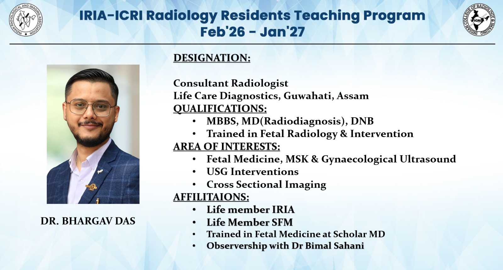
Official certification in advanced fetal ultrasound, high-risk obstetrics, and interventional fetal procedures.
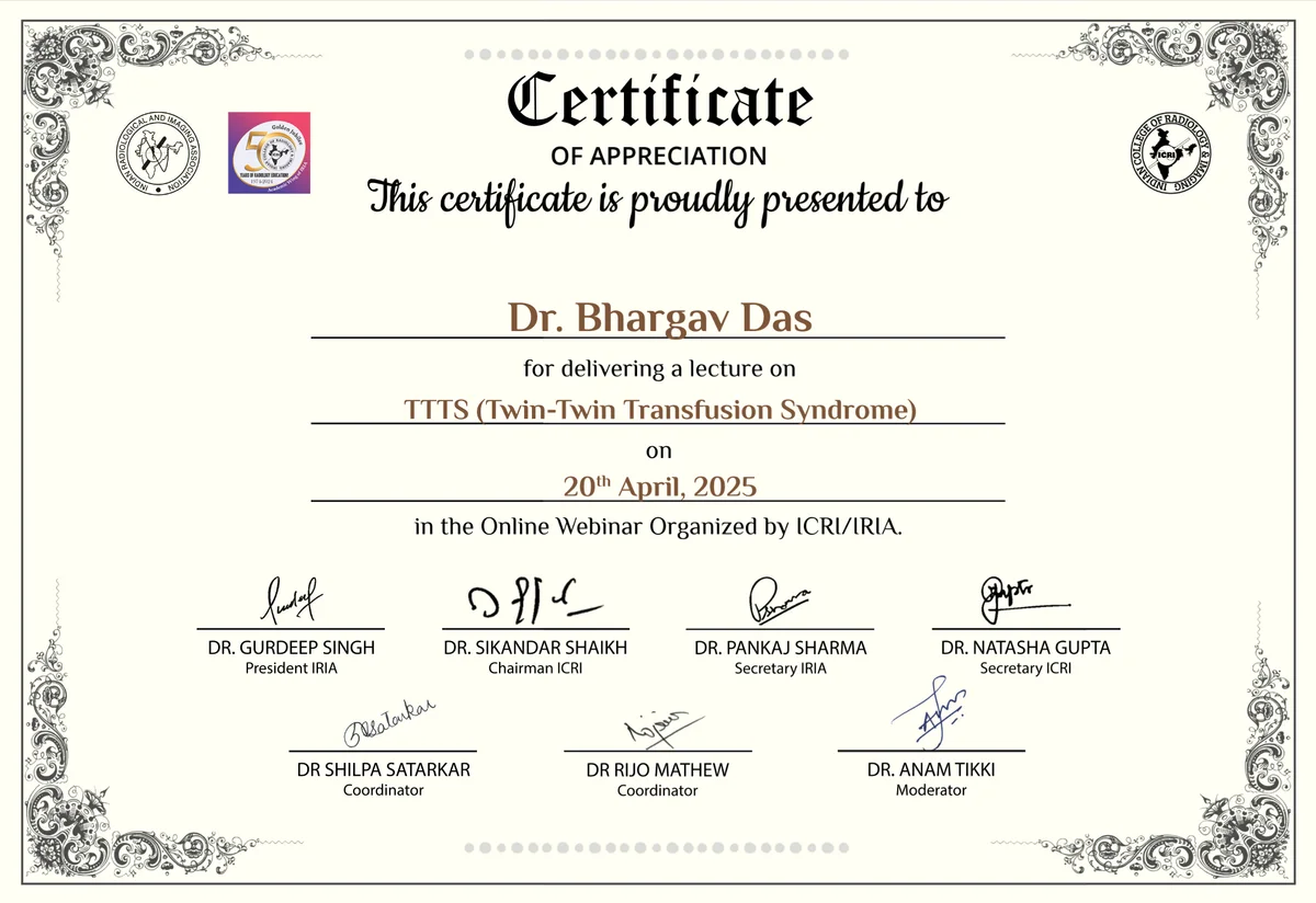
Diplomate certification issued by the National Board of Examinations, India.
Master of Medicine degree in Radiodiagnosis from accredited medical institution.
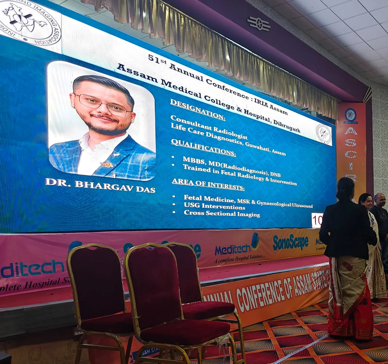
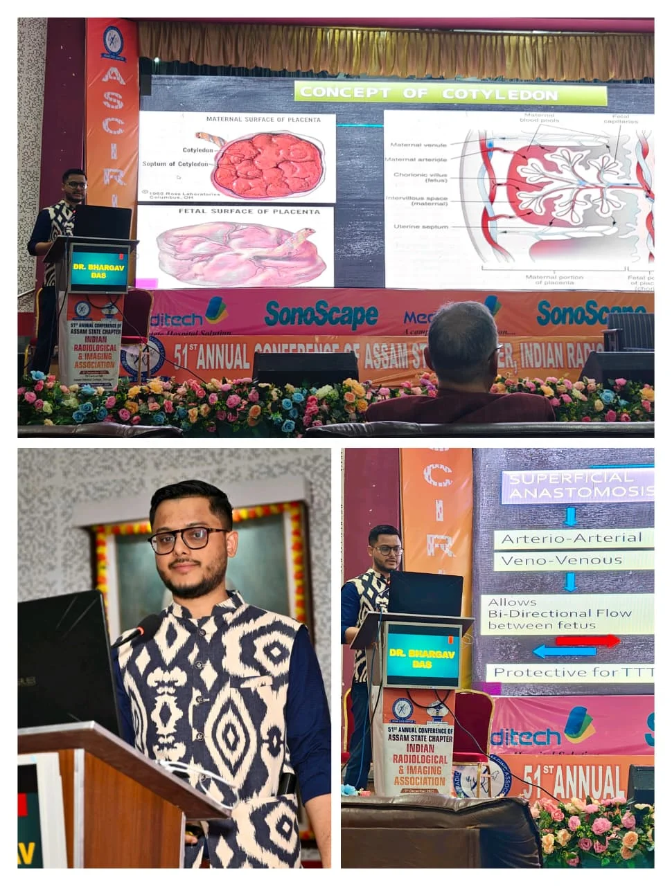
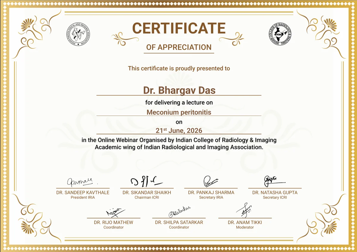
Dr. Bhargav Das actively participates in continuous medical education (CME), radiology conferences, multi-disciplinary team discussions, and academic peer interactions to stay at the forefront of modern diagnostic imaging standards.